What is a Cornea?
Cornea Defined
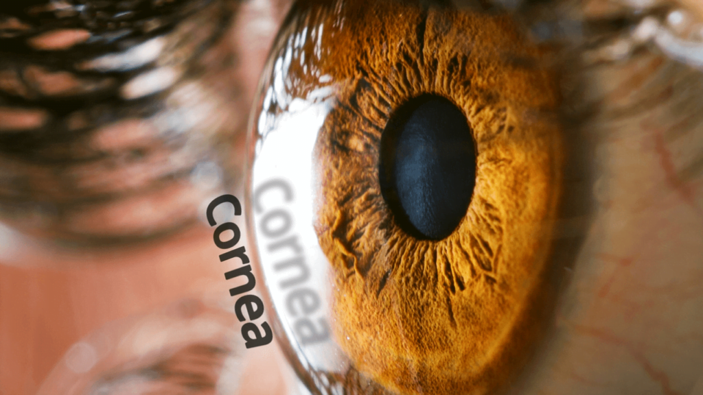
The clear dome over the iris (the color part of the eye) is called the cornea.
Another structure of the eye that the cornea is actually consistent with, the sclera, is also made up of hard collagen. Together, the cornea and sclera form the fibrous outer layer of the globe of the eye.
What makes the sclera and cornea different is the arrangement of the collagen fibers. The sclera is formed from very strong criss-crossing collagen tissue that gives the eye its shape, provides attachment for extraocular muscles, and protects the inner tissues of the eye from injury. This criss-crossing design of the collagen also makes the sclera white and opaque (light can not pass through it).
The cornea, on the other hand, is transparent because the collagen fibers are aligned in precise parallel layers and have a specific state of hydration that is maintained.
How the Cornea Got Its Name
The origin of the word “cornea” comes from the Greek word “keras,” which means “horn,” and the Latin word for horn which is “cornu”. Cornea got its name because a long time ago, people used to think that the cornea was made from the same material as animal horn’s keratin (notice the kera- part of keratin). Keratin is a tough fibrous protein that can be found in hair, fingernails, and skin. The cornea, however, is made up of a protein called collagen that is also a tough fibrous protein, but it is more commonly found in connective tissues like that which is found in tendons and ligaments.
The Cornea’s Role in Eyesight
Light enters the eye through the transparent cornea. Not only does the cornea serve as a “window’ for light to pass through, it also does the majority (65-75%) of the bending (refraction) of light to focus light onto the fovea, which lies in the center of the macula -the central vision zone of the retina – which lines the back of our eyes.
When light is focused perfectly on the fovea of a relaxed eye when distant parallel light enters the eye, the state of optics is referred to as emmetropia. When the cornea does not correctly bend the light to focus on the fovea, vision conditions occur.
- When light is focused at one point in front of the fovea, the optics is referred to as myopia (aka: nearsighted).
- When light is focused at one point behind the fovea, it is referred to as hyperopia or hypermetropia (aka: farsighted).
- When light is refracted to stretch focus across two points (usually because the cornea is flatter in one direction, and steeper in the perpendicular direction), it is called astigmatism.
Cells within the cornea are not nourished from blood with oxygen and food like all the other cells of our body. If blood flowed through the cornea, then the cornea would no longer be transparent. Instead, it would be red and opaque. Therefore, the cornea has to get its oxygen and nutrition from elsewhere.
Internally, aqueous humor provides this support with a constant flow of fluid that services the deeper layers of the cornea. Externally, the cornea gets its nutrition from the tears, and oxygen directly from the air around it. Oxygen actually diffuses from the air, into the water part of the tears, and then into the corneal tissue. At night, the cornea actually “steals” oxygen in a similar fashion from the back of our lids (that’s why lids are pink if you invert them – they are loaded with blood vessels meant to bring oxygen to a closed, or “sleeping”, eye).
How do Contact Lenses Work with the Cornea?
Corrective lenses with both glasses and contacts work similarly to realign the focus of light onto the fovea. Both glasses and contact lenses have different curves cut on the front surface as compared to the back surface to provide dioptric power (unit of measurement used for refracting light).
What is different between the two is the fact that contact lenses actually “contact” (hence the name “contact” lenses) the cornea. This “contacting” of the lens to the cornea is key to issues that must be addressed for contact lenses to be worn comfortably and safely.
Contact lenses placed on top of the cornea, therefore, disrupt both the tears and oxygen flow to the eye. A well designed contact lens will limit this tear and oxygen disruption to a minimum. Our tears have over 650 identified individual components, each with a different function that acts to lubricate, moisturize, nourish, protect, heal, oxygenate, and hydrate (to name a few) the cornea. Since a contact lens bathes in the tears of the cornea, often the balance of these tear components is at risk. Also, since contact lenses are a new barrier between the eye and its oxygen supply (the air), the contact must have a way to distribute oxygen through the lens itself and allow oxygenated tears to flow freely under the lens to limit oxygen deprivation of the cornea.
Years of contact lens technological advancements have served to make soft contact lenses much less problematic to the cornea than ever before. Oxygen permeability has increased significantly, especially with the use of silicone hydrogel materials in newer designs of soft lenses. Also, greater understanding of the components of tears have led to better contact lens designs that limit tear disruption or at least better support tear function in corneal health.
A properly worn contact lens is, overall, a safe form of correcting vision. It is improper care and poor wearing habits, such as overwearing and sleeping in contact lenses, that most often result in disruption to the eye’s natural flow and are the greatest risk of harming the cornea. Problems that arise with contact lenses are not only a threat to vision, but they can also be quite painful. The cornea is loaded with pain receptors so that even the slight issue of the cornea can be painfully perceived. Along with normal pain sensitivity, damage to the cornea will also commonly cause light sensitivity (aka: photophobia).
Common Vision Conditions that Involve the Cornea
Contact Lens Related Cornea Problems:
- Contact Lens Associated Red Eye (CLARE): often occurs, especially when contact lens wearing recommendations are ignored. The resultant problem causes the eye to appear red, feel irritated, and experience photophobia. Often the cornea swells and has an accumulation of inflammatory cells (infiltrates) that could develop into corneal ulcers (holes in the surface tissue of the cornea) and/or corneal scars that could result in vision loss if they occur on the visual axis
- Corneal Neovascularization: when too much oxygen flow is restricted from the cornea, the cornea will reach out to surrounding tissues to help bring oxygen into the cornea. If these new growing vessels get too close to the center of the cornea, vision disruption or loss can occur.
- Superficial Punctate Keratopathy (SPK): the top layers of the cornea (the epithelium) becomes irritated and damaged
- Keratitis: inflammation of the cornea
- Infection: bacteria, virus, fungus, or acanthamoeba growth can overwhelm the cornea if the defense mechanisms of the eye have become suppressed
- Infiltrates: accumulations of inflammatory cells that could lead to ulcers or scarring
- Ulcers: holes in the surface tissue that can develop due to infection of inflammation or both
- Edema: swelling with extra water that can disrupt normal layering of the collagen to the point where the cornea becomes “hazy” with a disruption clear optics
- Warpage: especially noticed with rigid contacts where the cornea conforms to the shape of the contact lens. This a disruption of the “natural” shape of the cornea, and often takes patience for restoration of its shape
Other Corneal Eye Conditions
- Foreign Bodies: tiny objects get stuck in the cornea, often from machines like grinding wheels, and frequently leave permanent scars
- Corneal Abrasion: a scratch to the cornea surface
- Recurrent Corneal Erosion: this may occur when deeper layers of the surface cornea are scratched, and the cornea heals, but does not fully heal. The surface of the cornea may stick to the lid during sleep, and like a piece of paper being pulled off of a lip, the sticky lid may reopen the previously scratched area of the cornea. The recurrence of the scratch can even occur many years after the original scratch, and has the tendency to “scratch/erode” over-and-over again.
- Anterior Basement Membrane Dystrophy (aka: Epithelial Basement Membrane Dystrophy): A genetic disorder of the a surface membranes of the cornea that can cause issues spontaneous “scratches” of the cornea upon opening the eyes from sleep, similar to the experience of a recurrent corneal erosion,
- Fuch’s Dystrophy: the inner layer of the cornea (the endothelium), is the cornea’s water pump. It constantly acts to pump water out of the cornea to maintain corneal hydration and the precise layering of collagen in the cornea for clear transparent tissues. When this genetic problem occurs, the pump begins to fail, and water backs up in the cornea, and can disrupt vision slightly or severely, depending on the amount of pump failure
- Dry Eye Syndrome: a deficiency in the normal tear layer that can lead to irritation, desiccation, inflammation, or infection of the cornea
- Meibomian Gland Dysfunction: the evaporative and more common form of dry eye created because the oil that usually mixes with the tear is not in high enough quantities, so the water of the tear dries. When the water dries, the cornea becomes irritated, so protective water flows to the rescue, but the oil can’t catch up… so the watery tear dries again.
- Aqueous Deficiency: the less common form of dry eye that occurs when the eyes do not produces enough water to maintain a stable tear and corneal health
- Pterygium: a growth of vascularized tissue from the conjunctiva (the transparent top layer of tissue that covers the sclera directly adjacent to the cornea) grows (usually in the 3:00 or 9:00 clock-face position) onto the cornea, and has potential to affect vision.
- Keratoconus: thinning of the cornea that causes the cornea to bulge and warp with the tendency to severely disrupt normal optics of the eye and glasses have limited effectiveness
Tips for Maintaining Eye Health of the Cornea
Wear Contact Lenses Safely:
- Wash your hands before handling
- Don’t sleep or nap in them
- Always clean and rinse them before and after insertion
- Let the eyes breathe without contact an hour before and after bedtime
- Throw them out at the recommended interval
- “When in Doubt, Take Them out”
Check out our other guides:
How to put contact lenses in →
How to remove contact lenses →
Let the Eyes Breathe
When contact lens issues arise, quite often the best answer is to remove the contact lens and let the eye breathe. When the natural oxygen flow and tear mechanisms are no longer disrupted, the eye often can restore order itself.
See an Eye Doctor
When you have eye problems, see an Eye Doctor, such as an Optometrist or an Ophthalmologist, to evaluate the eye issue. They have the best instruments and experience to evaluate, diagnose, and ultimately treat your eye conditions.
Artificial Tears
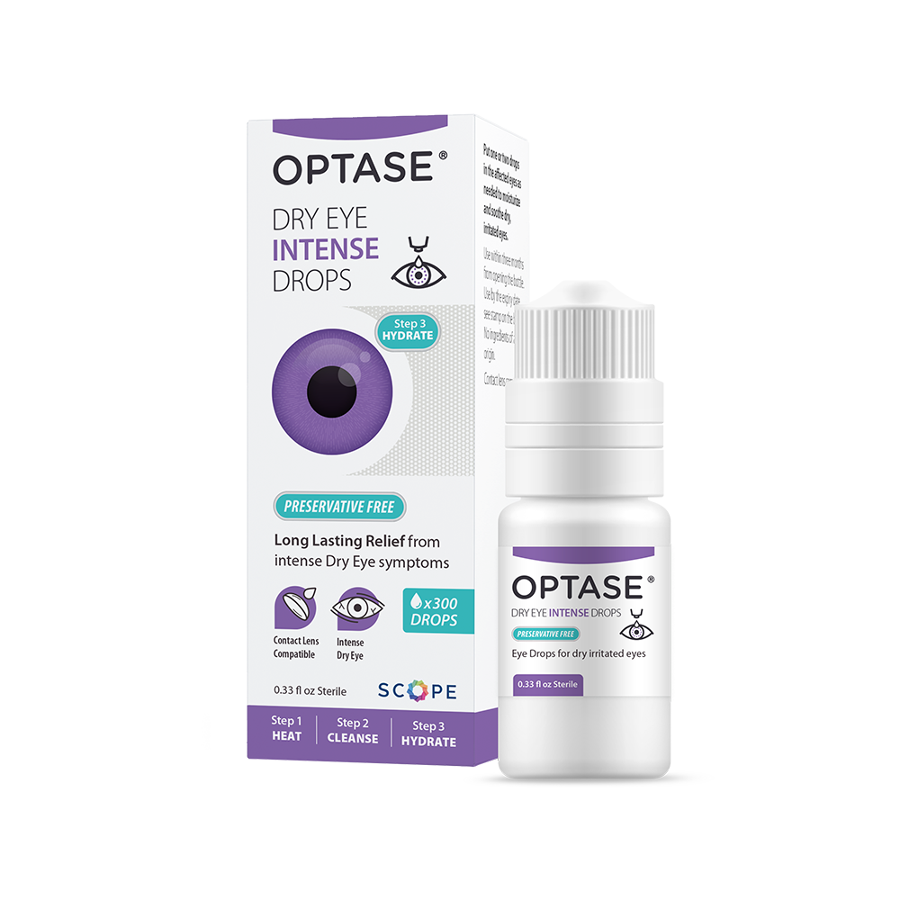
“Help you now”… when your cornea dries out due to poor tear oil disruption, the cornea will often become irritated and watery. Artificial tears stabilize the tear layer for about 10-20 minutes, and hopefully allow enough time for your natural oil from the meibomian glands of your eyelids to “catch back up” .
Nighttime Eye Ointments
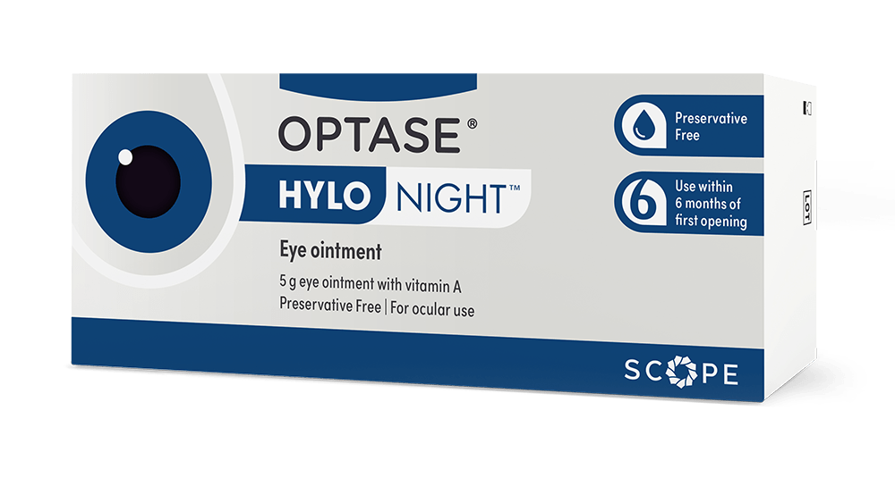
“Help you tomorrow”… at night, the oil glands of our eyelids go to sleep with us. Oil is not secreted like it is into our tears during sleep. This often leads to a drying of the cornea that becomes obvious upon waking, and is demonstrated by irritated and watery eyes. Nighttime eye ointment helps lock moisture on our corneas while we sleep, so instead of waking up with dry, irritated corneas, you may wake up with healed, moist corneas. It’s a great way to start the day.
Hot Compresses
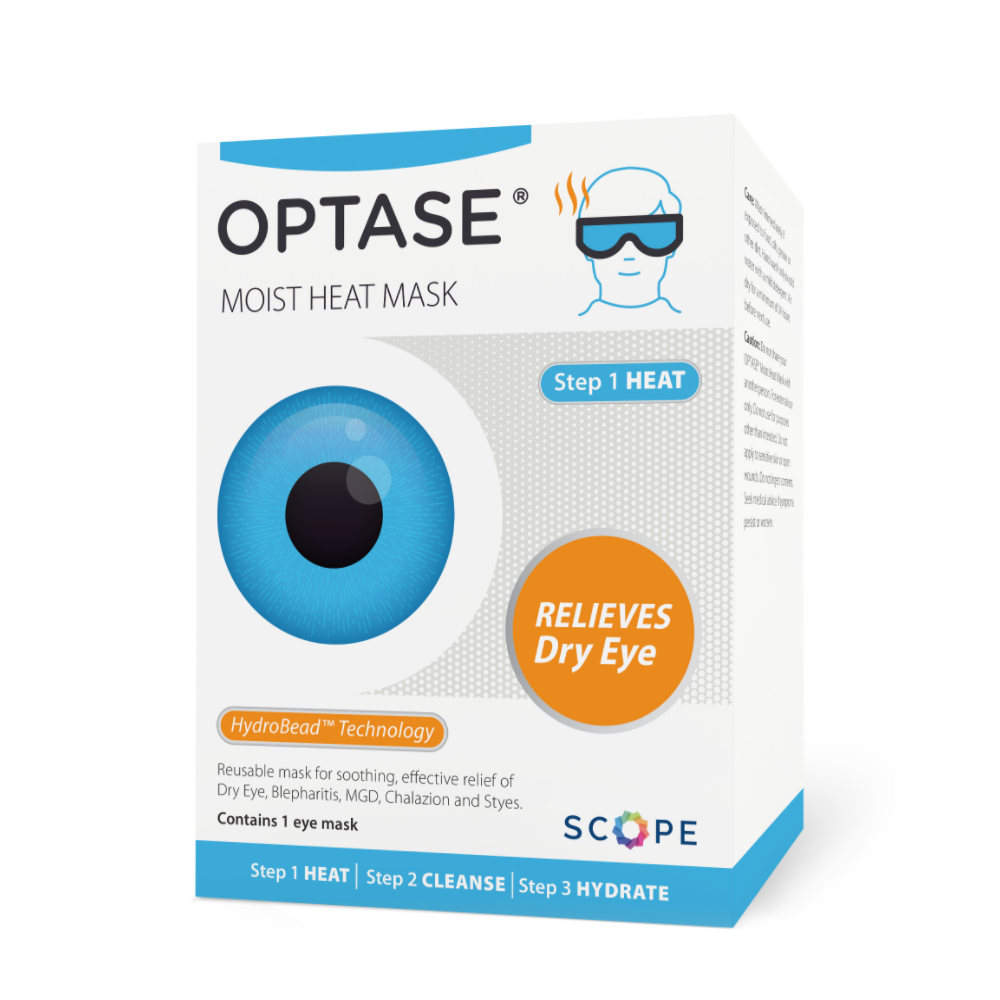
“Help you in the future”… Helps the cornea through the tear health mechanisms of heating the lid’s oil to create a smoother flow of tears and by increasing blood flow around the meibomian glands (oil glands) of the eyelids.
Omega-3 fatty acids
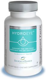
“Help you in the future”… Omega-3 may help increase the long term health of the eyelid’s meibomian oil glands and by increasing the blood flow around the meibomian glands, and by affecting the oil itself to flow more freely to limit dry eye symptoms that affect the tears and ultimately the cornea.
Punctal Plugs
These small plugs are inserted into the tiny holes (puncta) located in the top and bottom lid near the angle on the “nose-side” of each eye. By blocking these puncta, tears are stopped from entering the tear drainage system of the eye to decrease the outflow of tears. This method of dry eye therapy is used, most commonly, to treat the less common form of dry eye, Aqueous Deficiency.
Eye Protection
Wearing proper safety glasses, sunglasses, or regular glasses to protect your eyes from flying objects, tree branches, fingernails, UV light, and moving air (moving air increases tear evaporation) to name a few.
Eat Healthy, Drink Healthy, Be Healthy
A healthy body, leads to healthy eyes.
Need Consistanly Cheap Contacts?
DeliverContacts.com always guarantees you are paying low prices, every time you buy. We will never play games with our pricing or take part in manipulative discounts. Just consistently cheap contacts, forever.
Give your box a search below and see for yourself! 100% Free shipping and returns on all products!
