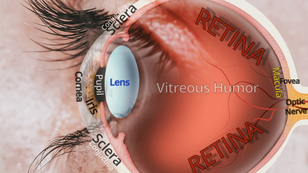
The Parts of Your Eye (Eye Anatomy)

Our body has five main senses:
- Vision
- Hearing
- Smell
- Taste
- Touch
The eye is the vision sensory organ. What actually perceives this sensory information, however, is the brain. All of our sense organs report to the brain for processing, understanding, considering, and responding to the information sent. The brain is also responsible for all of our thinking, remembering, moving, balancing, learning, speaking, and regulating nearly every part of our body.
But more than any other function of the body, more of the brain is dedicated to the eye than anything else. An amazing one-third to one-half of our usable brain is designed around the eye and vision. In fact, the eye actually has an entire lobe of the brain, the occipital lobe, devoted entirely to aspects of vision. This area is designated as the “visual cortex” and it is located in the back of our brain. Kind of weird, right? Our eyes, which are on the front of our head, course back to the back of our head to process visual information. So when you hear that someone has, “Eyes on the back of their head,”… their kind of right… in a way anyway.
The Eye as an Optical System
The eye perceives a certain part of the electromagnetic spectrum called the visible spectrum. The visible spectrum encompasses light wavelength size from 380-750 nanometers, which includes the various colors of the rainbow including Red, Orange, Yellow, Green, Indigo, and Violet (the popular mnemonic, ROY-G-BIV), and every possible combination of color that we see.
To perceive a detailed image, however, light has to be focused. In nature, light is divergent. That means that light spreads out infinitely in every direction from each point it emanates or is reflected until it is blocked or absorbed. This diverging light is noticed the closer you get to that point of light. Beyond twenty feet (six meters) from the source, for the most part mathematically, the diverging rays are parallel when they enter our eye, but they still need to be focused back to a single point to be seen clearly. And that is what the eye does. The eye can focus both parallel and diverging light back to a single point, and then send the information back to the brain for processing. The healthy eye uses about 125,000,000 points to decipher incoming light at every moment of our entire life. An amazing feat for such a small eye.
The External Parts of the Eye
Before light enters the eye, it must pass through the tears, a fluid that hydrates, lubricates, and protects the clear cornea. The tears and cornea are serviced by the eyelashes and eyelids to protect, clean, and produce these tears.
The cornea is the first structure that does the major bending of light (refraction of light) to refocus light to a single point. The clear cornea is surrounded by the white and opaque sclera which serves to block all other light from entering the eye, protect structures of the eye, and serve as an attachment point for the muscles that move the eye.
The sclera is covered by two layers: the conjunctiva, a clear surface tissue, and the clear episclera, a clear tissue that is between the conjunctiva and sclera.
From the cornea, light passes through aqueous humor in the anterior chamber of the eye, which is the space between the front of the iris (the part of the eye that gives our eye its ‘color”) and the cornea. In the center of the iris is a hole called the pupil. The pupil is an opening for light to enter the inner part of our eye.
The Internal Parts of the Eye
Immediately behind the pupil is a space, called the posterior chamber, which is a thin space from where aqueous humor flows to nourish tissues in the area of flow. After light passes through the pupil and through the posterior chamber, light then passes through the crystalline lens. This lens does not have as much refracting power as the cornea, but it can fine tune focus for when light becomes more divergent when looking at detail closer than twenty feet.
Beyond the lens is the vitreous body which is a clear gel that occupies the space of the inner eye. Ultimately, however, light gets focused on the brain’s neuronal component that extends into the eye, the retina, on a zone for central and color vision, called the macula, and more specifically a point for our most detailed sharpest viewing vision, called the fovea.
All of the light that is focused onto the retina stimulates photoreceptors (rods and cones), which then transfer this information out of the back of the eye along nerves. All of the separate nerves that exit the back of the eye combine to form a bundle of nerves called the optic nerve. The optic nerve then separates at the optic chiasm to form optic tracts which carry the information to the visual cortex in the occipital lobe in the back of the cerebrum.
Learn More about the Internal Parts of Your Eye
The Incredible Function of Vision
It is astounding how an eye can function to see, and then to consider how two eyes can receive separate images and then combine them into one binocular image within the brain is even more amazing. The sense of vision requires so many details to work together so perfectly, it is no wonder why so much of our brain must be devoted to its workings. When you can, take the time to explore the individual parts of the eye by following the highlighted words to our linked pages explaining each of them in greater detail.
Need Consistanly Cheap Contacts?
DeliverContacts.com always guarantees you are paying low prices, every time you buy. We will never play games with our pricing or take part in manipulative discounts. Just consistently cheap contacts, forever.
Give your box a search below and see for yourself! 100% Free shipping and returns on all products!
